生物细胞成像简介
Benefits
Live cell analyzer
The Digital Holographic Imager using transmission DHM® is a unique system for dynamic quantification of living cells processes. It is achieved in a non-perturbing way, thus allowing to investigate challenging biological processes. Its novel technology allows to measure single cells and gather population statistics for events occurring at the millisecond to multiple-days time-frame without the need of cell labeling.
Endogenous contrast
The DHM® advanced technology takes advantage of the natural endogenous contrast of cells to visualize and quantify them in a non-perturbing way without having to use interfering external dyes. This natural contrast comes from the thickness and intracellular refractive index of the cells and yield multiple information related to the morphology and cell content (protein, lipid content, etc.) of the monitored specimen.
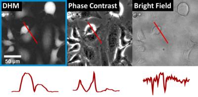
Digital Holographic Imaging technology
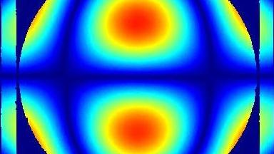
When light travels through a transparent object like living cells, its intensity is moderately altered thus only generating low contrast intensity images (like the one obtained with bright field microscopy) however the phase of the light is delayed by an amount proportional to the thickness and the refractive index of the cells.
Our patented DHM® technology allows to record in a single shot the phase information and obtain information related to a wide range of physiological parameters/biological processes (highlighted in the applications tab on the right).
Multimodality
DHM® can easily be combined with other modalities like fluorescence imaging of electrophysiological recording with our optional modules. Multimodality allows to combine synergetic information to achieved a better understanding of the biological processes monitored.
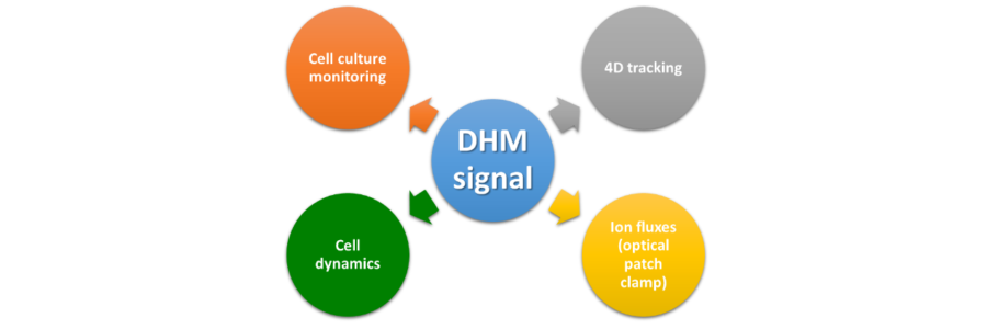
Parameters measured
Our CELL Analysis Tool software allows to easily extract all the rich information contained in the quantitative images related to:
- Morphology
- Intracellular content
- Dynamic parameters
- Population metrics
DHM® offers a unique quantitative phase signal that can offer multiple interpretations when investigating living cells.
A few key categories are highlighted in the other applications tabs.
Competitive advantages of DHM
DHM® offers key advantages when live samples need to be recorded in a non-perturbing way.
- Speed: microsecond acquisition
- Label-free: no marker needed, no phototoxicity (unlike fluorescence)
- Non-invasive: non-perturbing physiological measurements
- Quantitative: recordings proportional to physiological parameters
- High content analysis: large set of parameters acquired
- Perfect-focus: Off-line refocusing capability
- Ease of use: no sample preparation, direct imaging (no focusing or scanning)
These advantages combined allow to monitor living cells in real time for extended period of time. The absence of label allows for repeated measurement and even to perform an invasive measurement at the end of the DHM recording using another modality (for instance, fixation and antibody labeling).
Multimodality
The high content information provided by DHM can be correlated with simultaneous fluorescence measurements of the same field of view with the addition of the optional fluorescence module.