数字全息技术
专利技术
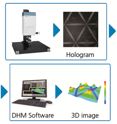
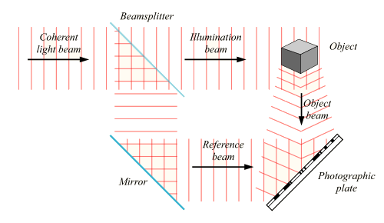
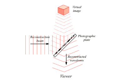
数字全息显微镜DHM® 是Lyncée Tec公司专利技术。数字全息技术使用CCD相机记录由参考光和物光干涉形成的全息图,再经由计算机进行数值运算后重建被测样品的三维图像,这一过程被称为“数字重建”。 DHM®的创新之处在于只需通过抓取单张图像既能获得样品的光学形貌信息,而抓取图像的过程是无须扫描的。另外,DHM®使用大量数值算法的方式在光学显微术中更是史无前例的。
数字全息显微镜DHM®的各种应用案例已经展示了这一款新概念显微镜对微观样品高精度超快速的三维成像功能,同时DHM®还具有了使用便利、通用性强、性价比高等优势。完整的方法描述请参考E. Cuche等人的综述文章: “Simultaneous amplitude-contrast and quantitative phase-contrast microscopy by numerical reconstruction of Fresnel off-axis holograms“
测试原理
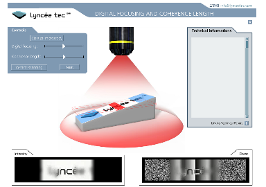
数字全息显微镜DHM® 是使用全息原理生产实时高分辨率的三维数字图像。全息图首先由参考光和物光相互干涉形成,再经由相机记录之后传输至电脑进行实时数字重建。 这个过程当中,一张全息图从获取到计算重建只需要几个微秒。 之后,数字全息显微镜DHM®专用采集分析软件通过分析计算获得的物体全息图,生成:
- 光强图:提供与传统显微镜一样对比度的图像
- 相位图:提供量化数值,对被测物体进行精确三维测量
在反射式数字全息显微镜中, 相位图以亚纳米精度揭示了被测物体的表面形貌。 在透射式数字全息显微镜中,相位图则展示了被测透明样品的相移信息,而这些数值对于特别是生物样品来说提供了重要的量化信息。 这种数字全息的方法使得视频和计算显微水平达到了一个前所未有的高度。
得益于数字全息的特殊原理,DHM®技术只需要使用软件补偿即可实现在传统显微镜中需要调整的各种光学参数,包括调整光学相差、数字图像聚焦、图像倾斜调整以及消除环境扰动等。
数字全息原理 (视频)
Holography is a well-established imaging technique. Since its discovery by Denis Gabor in 1947 [GAB48, GAB49, GAB51, GAB66] who was awarded the Nobel price in physics in 1971, large developments have been carried out. A description of the different forms and applications of classical holography is already developed in details in several books [GOO68, HAR96, COLL71, HAN79, STR69, SMI69, FRA87]. Furthermore, an exhaustive list of historical papers and an interesting overview of the developments of holography from its discovery to the present can be found in E.N. Leith’s overview [LEITH97].
It is however important to mention that even if digital holography took a great importance in the last years, holography progresses in non digital or more classical holography cannot be reduced to 3D spectacular “art images”. Research launched in the 50s and 60s by several famous personalities as D. Gabor, E.N. Leith, A. Lohmann, R.J. Collier, J. Upatnieks, G. Stroke, N. Hartman, Yu. N. Denisyuk, S. Benton, R.F. Vanligte, J.W. Goodman, R. Dändliker and N. Abramson, emerge onto a lot of different applications such as holographic data storage [SHEL97, ORT03], photorefractive crystals hologram capture [ROO03], light-in-flight for ultrafast phenomena recording [YAMA05], metrology applications, TV holography [POO05] and so on. Furthermore, all aspects of holography is a source of inspiration for the development of digital holography. For example, the configuration for DHM presented by E. Cuche [CUC99b] and used in this work was presented for the first time in 1966 by R.F. VanLighten and H. Oserberg [VAN66].
Concerning digital holography, the first computer reconstructions of holograms go back to the late sixties, some twenty years after the publication of Gabor’s landmark papers. The idea was proposed for the first time in 1967 by J.W. Goodman and R.W. Laurence [GOO67]. Numerical hologram reconstructions were initiated in the early 1970s by M.A. Kronrod and L.P. Yaroslavsky [KRO72a, KRO72b]. Nevertheless, the holograms were still recorded on a photographic plate, developed, optically enlarged and finally sampled before being numerically reconstructed.
A complete digital holographic set-up in sense of digital recording and reconstruction was achieved first by Coquoz et al. [COQ92, COQ93a, COQ93b] followed by U. Schnars and W. Jüptner in 1994 [SCHN94a]. The introduction of a coupled charged device (CCD) camera to record Fresnel holograms suppresses the recording on media such as photographic plate, photopolymers and photorefractives. It allows faster acquisition and reconstruction rates, as well as larger flexibility.
Important steps in the evolution of the technique and algorithms have been: the acquisition through endoscopic device [COQ95, SCHE99, SCHE01, KOLE03, PED03], the use of high wavelength [ALL03], and short-coherence laser source [CUC97, IND00, PED01a, PED02, MAS05, MAL05]. A very important step was the retrieval of the phase information in addition to the amplitude. Different techniques exist to reconstruct the phase. In-line techniques require phase-shifting procedures performed either by several successive hologram acquisitions [YAM97, ZHA98, LAI00, GUO02, YAM03, AWA04, MILGA05] or by simultaneous acquisitions [KOLI92, MILL01, WYA03]. In off-axis configurations, U. Schnars [SCHN94b] has demonstrated the possibility of measuring specimen deformations by evaluating the phase difference between two states of the specimen. However, this double exposure technique does not give the absolute phase of the specimen. A solution for absolute phase measurements has been proposed by E. Cuche in off-axis geometry [CUC99a] and delivered patent [CUCPat]. A flat reference surface is taken on a flat part of the specimen and a procedure is performed to compensate for the phase deformation. The complete wavefront is thus reconstructed out of a single hologram, which retrieves the initial aim of holography. E. Cuche also showed [CUC99b] that the phase compensation technique could be applied in holographic microscopic methods. Later on other techniques were developed to perform the phase reconstruction out of a single hologram [LIE03, LIE04]. P. Ferraro [FERR03a] performs a method in which a reference hologram is recorded and then subtracted to the hologram of interest to compensate for the phase deformations. G. Indebetouw developed a method, called Spatiotemporal digital holograph, that avoids the need for high spatial bandwidth detectors and high spatial coherence, leading to speckle noise [IND99, IND01]. Nowadays, digital holographic microscopy has become a wide used method [ZHA98, TAK99, TIS01, XU01, YAM01, DUB02, CARL04, COP04].
Other developments include color digital holography [KAT02, YAM02, ALM04, JAV05b], polarization digital holography [LOH65, COLO02b, COLO04, COLO05], synthetic wavelength digital holography [ONO98, WAG00, GAS03], tomography [KIM99, KIM00, DAK03, MONT06], optical diffraction tomography [CHAR06a, CHAR06b] and several aberration compensation techniques [CUC99b, STA00, PED01b, DEN02, FERR03b, FERR03a, IND01, MAL05, YON05, COLO06a, COLO06b, COLO06c]. The development of DHM allows truly non-invasive examination of biological specimens and therefore it becomes an increasing powerful technique for biomedical applications [COLO02a, TIS05, MAS05, MAL05, MARQ05, JEO05, JAV05a, RAP05].